Entry Version:
Citation:
Pancreapedia: Exocrine Pancreas Knowledge Base, DOI: 10.3998/panc.2013.16
| Attachment | Size |
|---|---|
| 421.4 KB |
1. Introduction
Autoimmune pancreatitis (AIP) is a newly recognized entity of pancreatitis that can mimic malignancy (6). Patients with AIP and pancreatic cancer have many clinical features in common, such as a higher prevalence among elderly males, frequent presentation with painless jaundice, development of diabetes mellitus, and elevated levels of serum tumor markers. Radiologically, focal swelling of the pancreas, the “double-duct sign” (representing dilation of both biliary and pancreatic ducts), and encasement of peripancreatic arteries and portal veins can be seen in both AIP and pancreatic cancer (3, 5). Since AIP responds dramatically to steroid therapy, differentiation of AIP from pancreatic cancer is of paramount importance to avoid unnecessary laparotomy or pancreatic resection. As definite serological markers for AIP are lacking, diagnosis of AIP is currently based on a combination of clinical, laboratory, and imaging studies. Imaging of the pancreatic duct with endoscopic retrograde pancreatocholangiography (ERCP) plays an important role in the diagnosis of AIP.
2. Autoimmune pancreatitis and ERCP
The concept of AIP emerged from the pancreatographic study of chronic pancreatitis. Four cases of peculiar pancreatitis showing diffuse irregular narrowing of the entire main pancreatic duct (MPD) on endoscopic retrograde pancreatography (ERP) were reported by Toki et al. of Tokyo Women’s Medical University in 1992 (14). Yoshida et al., from the same group, proposed the concept of AIP on the basis of a case with diffuse irregular narrowing of the MPD on ERP that was steroid responsive (15). Pancreatographic findings of diffuse or segmental irregular narrowing of the MPD on ERP were mandatory in the Japanese clinical diagnostic criteria for AIP in 2006 (9).
ERCP features suggesting autoimmune pancreatitis
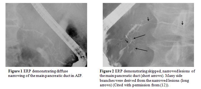
Unlike obstruction or stenosis, narrowing of the MPD (in which the duct diameter is smaller than normal with irregular walls) is seen in AIP patients. Typical AIP demonstrates narrowing for more than one-third of the entire length of the MPD. Diffuse irregular narrowing of the MPD, in which the length of the MPD involved is more than two-thirds of the entire MPD, is rather specific to AIP (Figure 1) (10). In one study (that compared ERCP findings of 48 AIP patients and143 pancreatic cancer patients), the length of the narrowed portion of the MPD was significantly longer, and the diameter of upstream MPD was significantly smaller in AIP than in pancreatic cancer (13).


Furthermore, pancreatographic findings such as the lack of MPD obstruction, skip lesions in the MPD (Figure 2), side branch derivation from the narrowed portion of the MPD (Figure 2), narrowed portion of the MPD >3-cm-long, and maximal diameter of the upstream MPD <5 mm, were highly suggestive of AIP rather than pancreatic cancer (Table 1).
Although stenosis of the lower bile duct is frequently detected in both AIP and pancreatic cancer on cholangiography, stenosis of the intrahepatic or hilar bile duct is seen only in AIP patients. Differentiating a short narrowing of the MPD in AIP from stenosis in pancreatic cancer is difficult (Figure 3), and some cases of pancreatic cancer have pancreatographic findings similar to those of AIP (8). According to an international multicenter study, the presence of pancreatic duct strictures (single or multiple) without upstream dilatation (<5 mm) offered the highest specificity for AIP (>90%) (12).
Histopathological examination of type 1 AIP shows lymphoplasmacytic sclerosing pancreatitis (LPSP), characterized by dense lymphoplasmacytic infiltration and fibrosis in the pancreas. Abundant lymphoplasmacytic cells infiltrate with fibrosis around interlobular pancreatic ducts including the MPD. Although the periductal inflammation is usually extensive and distributed throughout the entire pancreas, the degree and extent of the inflammation differ from duct to duct depending on the location of the pancreas involved. The infiltrate is primarily subepithelial and inflammatory cells rarely infiltrate the epithelium. It encompasses the pancreatic ducts and narrows their lumen (1, 4, 7). On the other hand, pancreatic cancer cells infiltrate scirrhously, destroying the epithelium of the pancreatic and bile ducts, and frequently obstruct the MPD and branch pancreatic ducts. These histopathological differences around the ducts may account for the different pancreatographic findings between AIP and pancreatic cancer.
ERCP for the diagnosis of autoimmune pancreatitis: variation in usage worldwide
There are several specific features of ERCP that appear potentially useful for differentiating AIP from pancreatic cancer. However, local expertise and patterns of practice in the use of various tests vary considerably worldwide. Although diagnostic ERP is frequently performed in Japan, Western endoscopists generally avoid injecting the pancreatic duct in patients with obstructive jaundice for fear of causing pancreatitis. Instead, AIP is often diagnosed without an ERP in Western countries (2, 12).
According to the International Consensus Diagnostic Criteria patients with diffuse enlargement of the pancreas with elevated serum IgG4 levels can be diagnosed with AIP without pancreatography (Diagnosis of Autoimmune Pancreatitis) (11). However, based on the high specificity for AIP, the presence of a long (>1/3 length of the MPD) or multiple strictures without marked upstream dilation is strong collateral evidence for diagnosing AIP in those with atypical parenchymal imaging, such as segmental or focal pancreatic enlargement. Thus, ERP can be helpful in differentiating AIP from pancreatic cancer, and in these casesbrush cytology of the narrowed portion of the MPD is often performed.
Role of MRCP in the diagnosis of autoimmune pancreatitis
Magnetic resonance cholangiopancreatography (MRCP) has become a popular non-invasive method for obtaining high-quality images of the pancreaticobiliary tree, and is replacing diagnostic ERCP for the diagnosis and follow-up of many pancreatobiliary diseases. However, since the narrowed portion of the MPD seen on ERCP in AIP can rarely be visualized on MRCP due to the inferior resolution of MRCP, this modality cannot yet replace ERCP in the diagnosis of AIP. However, MRCP findings such as skipped narrowing of the MPD with lack of upstream MPD dilatation suggest AIP. Furthermore, as resolution of the pancreatic and bile ducts after steroid therapy can be fully evaluated on MRCP, this approach is useful to determine the effect of steroid therapy and follow up after steroid therapy (13).
3. Summary
When utilized, ERP can demonstrate specific findings for AIP including long or multiple strictures without marked upstream dilation of the main pancreatic duct. These findings can be particularly useful for differentiating from pancreatic cancer in those with atypical features of AIP on parenchymal imaging. Although MRCP may be useful to evaluate the effect of steroid therapy, the sensitivity for detecting changes in the main pancreatic duct of AIP is inadequate to replace ERP.
Acknowledgement
This work was supported in part by the Research Committee of Intractable Pancreatic Diseases, provided by the Ministry of Health, Labour, and Welfare of Japan.
4. References
- Chandan VS, Iacobuzio-Donahue C, and Abraham SC. Patchy distribution of pathologic abnormalities in autoimmune pancreatitis: implications for preoperative diagnosis. Am J Surg Pathol 32: 1762-1769, 2008. PMID: 18779731
- Chari ST, Takahashi N, Levy MJ, Smyrk TC, Clain JE, Pearson RK, Petersen BT, Topazian MA, and Vege SS. A diagnostic strategy to distinguish autoimmune pancreatitis from pancreatic cancer. Clin Gastroenterol Hepatol 7: 1097-1103, 2009. PMID: 19410017
- Kamisawa T, Egawa N, Nakajima H, Tsuruta K, Okamoto A, and Kamata N. Clinical difficulties in the differentiation of autoimmune pancreatitis and pancreatic carcinoma. Am J Gastroenterol 98: 2694-2699, 2003. PMID: 14687819
- Kamisawa T, Funata N, Hayashi Y, Tsuruta K, Okamoto A, Amemiya K, Egawa N, and Nakajima H. Close relationship between autoimmune pancreatitis and multifocal fibrosclerosis. Gut 52: 683-687, 2003. PMID: 12692053
- Kamisawa T, Imai M, Yui Chen P, Tu Y, Egawa N, Tsuruta K, Okamoto A, Suzuki M, and Kamata N. Strategy for differentiating autoimmune pancreatitis from pancreatic cancer. Pancreas 37: e62-67, 2008. PMID: 18815540
- Kamisawa T, Takuma K, Egawa N, Tsuruta K, and Sasaki T. Autoimmune pancreatitis and IgG4-related sclerosing disease. Nat Rev Gastroenterol Hepatol 7: 401-409, 2010. PMID: 20548323
- Kloppel G, Sipos B, Zamboni G, Kojima M, and Morohoshi T. Autoimmune pancreatitis: histo- and immunopathological features. J Gastroenterol 42 Suppl 18: 28-31, 2007. PMID: 17520220
- Nishino T, Oyama H, Toki F, and Shiratori K. Differentiation between autoimmune pancreatitis and pancreatic carcinoma based on endoscopic retrograde cholangiopancreatography findings. J Gastroenterol 45: 988-996, 2010. PMID: 20396913
- Okazaki K, Kawa S, Kamisawa T, Naruse S, Tanaka S, Nishimori I, Ohara H, Ito T, Kiriyama S, Inui K, Shimosegawa T, Koizumi M, Suda K, Shiratori K, Yamaguchi K, Yamaguchi T, Sugiyama M, and Otsuki M. Clinical diagnostic criteria of autoimmune pancreatitis: revised proposal. J Gastroenterol 41: 626-631, 2006. PMID: 16932998
- Okazaki K, Kawa S, Kamisawa T, Shimosegawa T, and Tanaka M. Japanese consensus guidelines for management of autoimmune pancreatitis: I. Concept and diagnosis of autoimmune pancreatitis. J Gastroenterol 45: 249-265, 2010. PMID: 20084528
- Shimosegawa T, Chari ST, Frulloni L, Kamisawa T, Kawa S, Mino-Kenudson M, Kim MH, Kloppel G, Lerch MM, Lohr M, Notohara K, Okazaki K, Schneider A, and Zhang L. International consensus diagnostic criteria for autoimmune pancreatitis: guidelines of the International Association of Pancreatology. Pancreas 40: 352-358, 2011. PMID: 21412117
- Sugumar A, Levy MJ, Kamisawa T, Webster GJ, Kim MH, Enders F, Amin Z, Baron TH, Chapman MH, Church NI, Clain JE, Egawa N, Johnson GJ, Okazaki K, Pearson RK, Pereira SP, Petersen BT, Read S, Sah RP, Sandanayake NS, Takahashi N, Topazian MD, Uchida K, Vege SS, and Chari ST. Endoscopic retrograde pancreatography criteria to diagnose autoimmune pancreatitis: an international multicentre study. Gut 60: 666-670, 2011. PMID: 21131631
- Takuma K, Kamisawa T, Tabata T, Inaba Y, Egawa N, and Igarashi Y. Utility of pancreatography for diagnosing autoimmune pancreatitis. World J Gastroenterol 17: 2332-2337, 2011. PMID: 21633599
- Toki F KT, Oi I, Nakasako T, Suzuki M, Hanyu F. An unusual type of chronic pancreatitis showing diffuse irregular narrowing of the entire main pancreatic duct on ERCP - a report of four cases. Endoscopy 24: 640, 1992.
- Yoshida K, Toki F, Takeuchi T, Watanabe S, Shiratori K, and Hayashi N. Chronic pancreatitis caused by an autoimmune abnormality. Proposal of the concept of autoimmune pancreatitis. Dig Dis Sci 40: 1561-1568, 1995. PMID: 7628283


